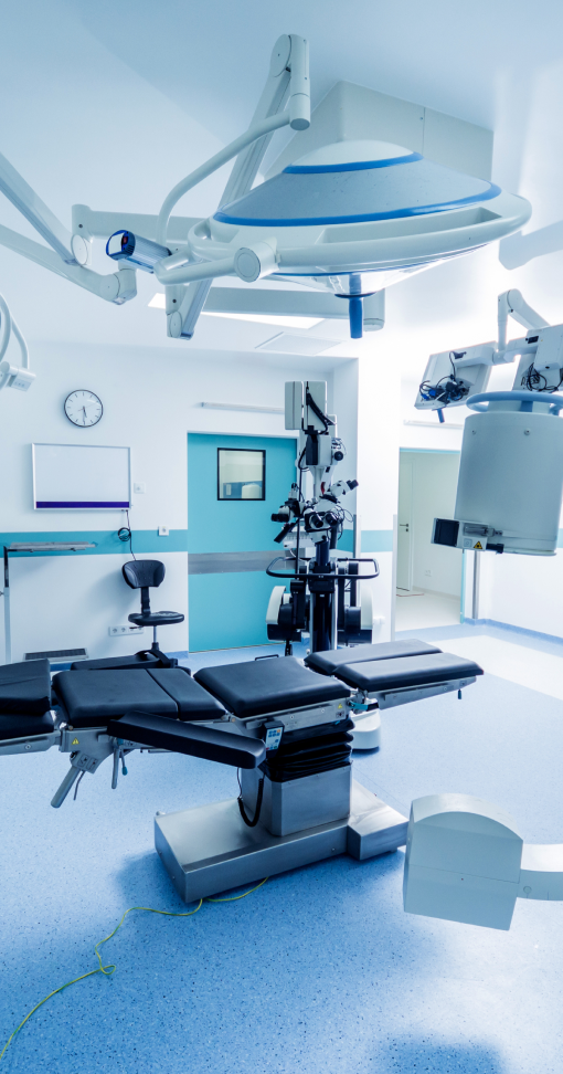
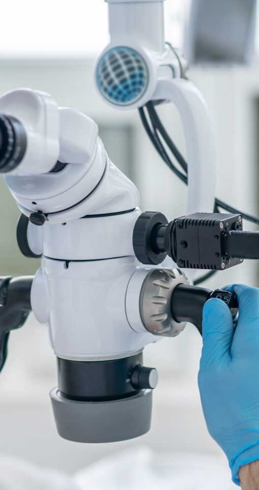
Partners & Vendors
We are thankful for all of our working relationships, and hope to add you to our list soon!
Our Latest and Most Popular Videos
Infantile hemangiomas are vascular tumors composed of proliferating endothelial cells. They uniquely undergo rapid expansion from birth to 6-8 months of age and subsequent slow dissolution over several years thereafter. Some hemangiomas are at risk of causing functional problems during their growth phase as seen in this upper eyebrow lesion obstructing the visual axis. Laser, surgical and medical treatment options are available for problematic hemangiomas. This patient was elected to undergo excision to completely remove the lesion and forego a long course of medical therapy (propranolol). Because of the their vascular nature, excision of hemangiomas requires careful planning and hemostasis. The hemangioma is marked in elliptical fashion along natural aesthetic facial lines along the brow. The inferior mark in incised first. Careful subdermal dissection is critical to completely excise to the hemangioma near the surface and find the appropriate plane. Control of bleeding is maintained by monopolar and bipolar electrocautery as well as dissecting the lesion from one side and alternating to the other. The plane of deep dissection is rarely below the subcutaneous layer thus protecting important nerves and vessels. Complete removal is possible. Closure is performed with dissolvable monocryl or PDS suture with dermabond superficially. A plastic eyeshield (blue) is placed at the beginning of case to protect the patient's cornea during the procedure. DOI: http://dx.doi.org/10.17797/zlvhux8afu
Watch the Full VideoThe following video depicts the excision of a 2nd branchial cleft fistula in a 12-month-old male. Fibrin glue dyed with methylene blue was used to assist with following the fistula tract to ensure complete excision of the lesion. Branchial cleft anomalies include fistulae, sinuses and cysts and most commonly occur in the lateral neck arising from the second branchial cleft. The patient may be completely asymptomatic, mildly affected or continuously impacted by the lesion. Recurrent inflammation, infections, drainage, and pain are common symptoms associated with these congenital anomalies. Surgery is the standard of care and recommended to alleviate symptoms, but recurrence rates are high, particularly if excision is incomplete. Methylene blue has been used to assist with complete excision of these lesions, but has several key drawbacks including spillage into nearby tissues, incompletely highlighting the lesion, and making the pathological examination more challenging due to significant tissue staining. The addition of fibrin glue to the methylene blue enables for the lesion to be well visualized with the dye without spilling into the adjacent tissue, and thereby reducing the risk of damaging nearby structures. The mixture also allows for efficient pathological examination for correct post-operative confirmation of the diagnosis.
Watch the Full VideoThe procedure in this video demonstrates the removal and replacement of a malpositioned Jones tube with endoscopic endonasal guidance to ensure proper tube placement within the nose.
Watch the Full VideoBilateral ear keloid excision with steroid injection. DOI# 10.17797/rfealpdd24
Watch the Full VideoThis video shows a pediatric patient with Van der Woude syndrome. He has lip pits that are classic for this syndrome and his family desired surgical correction. This video outlines and shows the steps of the modified simple excision technique as well as discussing tips for a successful surgery.
Watch the Full VideoMyringotomy with tympanostomy tube insertion is among the most common pediatric operative procedures and is indicated to provide ventilation of the middle ear. Surgical incision in the tympanic membrane (myringotomy) is followed by tympanostomy tube insertion to prevent premature closure of the incision site. The goal of the procedure is to reduce the frequency, duration, and severity of subsequent otitis media episodes and to prevent recurrence of middle ear effusions. Soham Roy (University of Texas at Houston Medical School) Thomas Mitchell (University of Texas at Houston Medical School)
Watch the Full VideoTitle: Nasopharyngeal Papillomatosis- A combined trans nasal transoral coblation assisted approach Authors - 1. Dr Deepa Shivnani- corresponding author MBBS, DNB Otolaryngology , MNAMS, Fellowship in Pediatric Otolaryngology Children’s Airway & Swallowing Center Manipal Hospital, Bangalore , India email- deepa.shivnani14@gmail.com 2. Dr E V Raman MBBS, DLO , MS Otorhinolaryngology Children’s Airway & Swallowing Center Manipal Hospital, Bangalore Here I am presenting a case of 16 yrs old boy, who had nasal block and occasional cough. Nasal endoscopy revealed an exophytic papillomatous growth in the nasopharynx. MRI showed lesion arising from the nasopharyngeal surface of the soft palate. The lesion was free from the posterior pharyngeal wall. The patient was taken up for the procedure under general anaesthesia. The transoral approach was followed first. The tissue was taken for histopathological examination followed by a traction suture placed over uvula for better visualisation. Once exposed, coblation device was used transorally with 45 degree hopkins rod transorally. The tissue was ablated with coblation and coagulation settings of 9:5 respectively. The base was ablated too, to prevent further recurrence. Tonsillar pillar retractor was then used for better visualisation and exposure. The coblation was then continued. The tissue was removed transorally as much as possible then trans nasal approach was performed. Then, the same coblation device with the same setting was used but the nasal endoscope was changed to O degree Pediatric scope due to space constraints. The lesion was pushed upward with the help of yankaurs suction tip for better exposure and the remaining tissue was removed with the help of same coblation device. The lesion was excised completely and successfully with minimal blood loss. The operative area was confirmed with the 70Degree hopkins rod for complete removal of the lesion. Post operative recovery was uneventful. Combined transoral trans nasal coblation assisted approach is potential to be safer, easier and less invasive than uvulo palato pharyngoplasty in Pediatric age group specially, in the areas which are difficult to access like nasopharyngeal surface of the soft palate what we showed in this video.
Watch the Full VideoContributors: James Kee In this video, we show the open surgical release of the A1 pulley to restore movement and alleviate triggering in a patient with stenosing tenosynovitis, or trigger finger. DOI #: https://doi.org/10.17797/punju11l92
Watch the Full VideoThis video demonstrates punctal dilation and insertion of a Mini-Monoka stent for treatment of epiphora due to punctal/canalicular stenosis.
Watch the Full VideoThe patient had an unidentified dermal filler placed outside of the United States over a decade ago. She developed a subsequent severe reaction which left her with extensive subdermal fibrosis and epidermal necrosis. Pathologic analysis revealed almost entire replacement of the dermal-epidermal layer with a foreign body and granulomatous reaction. The location at the cheek lower lid junction and the available lateral skin laxity deemed the rhomboid flap as the best option for reconstruction. Editor Recruited By: Michael Golinko, MD
Watch the Full VideoBecome an Industry Sponsor!

CSurgeries provides industry partners with direct access to members through our video channels. Viewers and subscribers will have the ability to watch the videos and access any tools and resources provided by any specific industry partner

CSurgeries provides a platform for industry partners to curate and share content to reach target surgeons and promote their products/events

Industry Partners demonstrate a commitment to professional development by providing an avenue for peer-reviewed video publication to their surgeons and/or members. Surgeons can submit CElite Videos for possible PubMed indexing

CSurgeries Partners receive brand recognition as a socially responsible supporter of the academic surgical community and surgical learning programs through promotion on our various marketing and communications channels