The leading global platform for
peer-reviewed surgical videos
Explore a collection of 500+ peer-reviewed surgical videos
from 300 contributors from around the world.
Trusted By:
Our Latest and Most Popular Videos
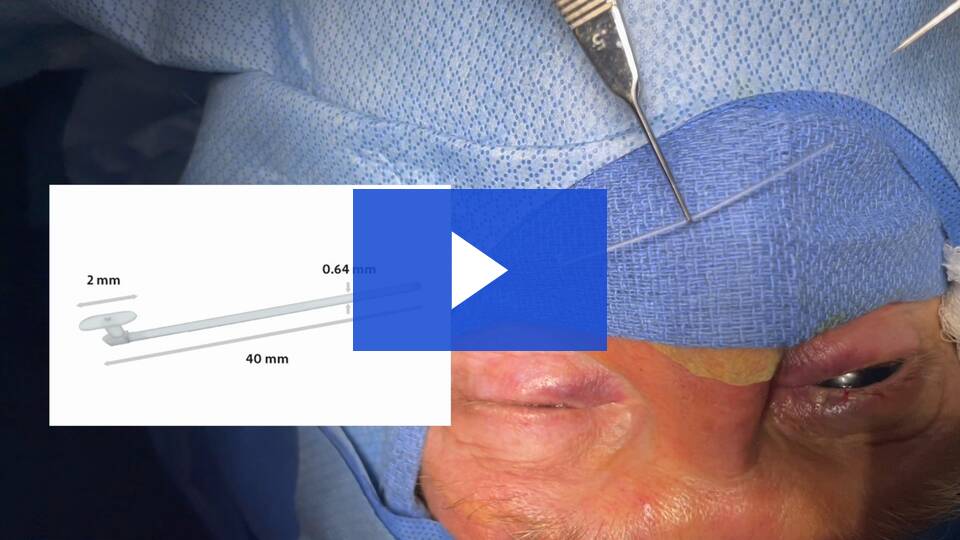
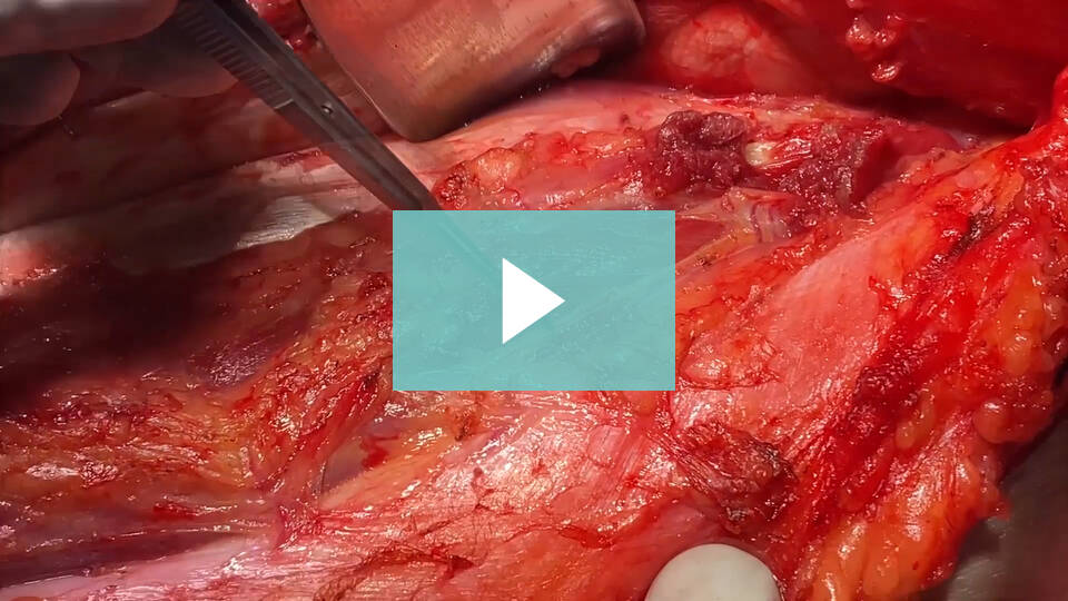
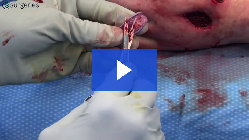
This video depicts the surgical excision of a pyogenic granuloma on the lower eyelid. The patient was brought into the operating room and general anesthesia was induced, the base of the lesion was injected with lidocaine and epinephrine. A warm compress was applied to soften the crusted lesion. The crust was then gently elevated revealing the giant pyogenic granuloma. A chalazion curette was used to completely remove the lesion. A chalazion clamp was utilized to stabilize the lid and for hemostasis purposes.
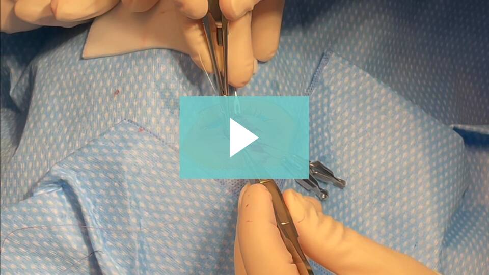
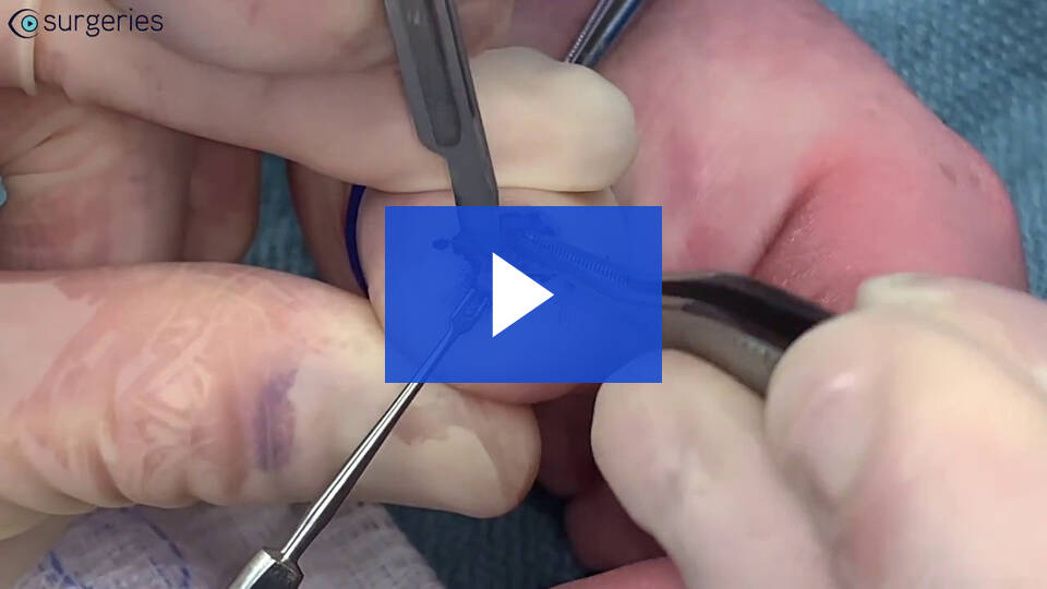
Watch the Full VideoThis video provides an elucidation of the surgical steps involved in performing an endoscopic perctaneous suture laterlization in a neonate with bilateral vocal fold paralysis.
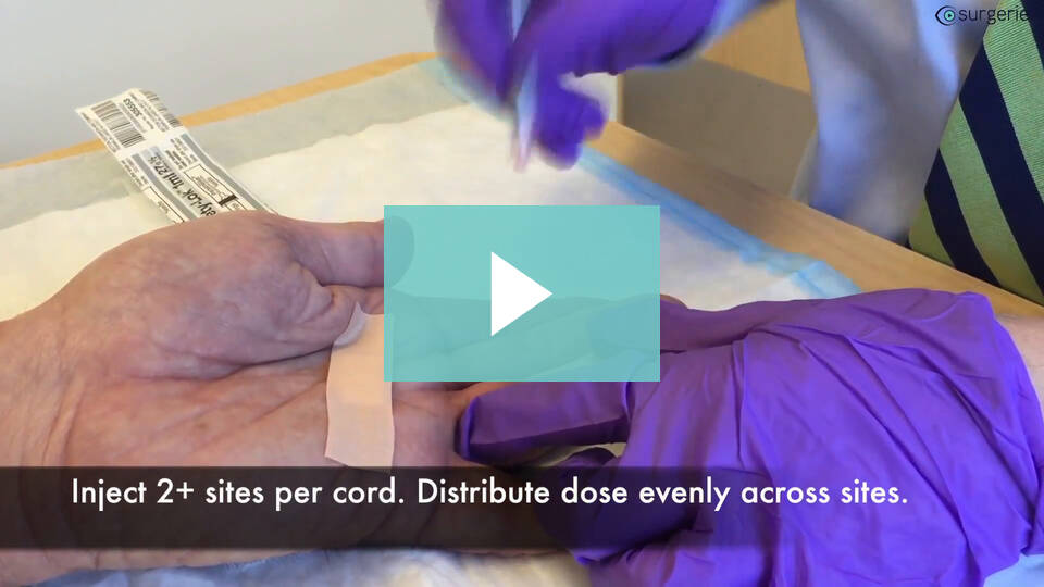
Watch the Full VideoFour Gland Duct Ligation with Botulinum Injections Background: This video visualizes the four-duct ligation surgery for chronic sialorrhea. Sialorrhea is characterized by the improper spilling of saliva, most commonly due to poor muscle coordination1. Controlling oral secretions with the perioral muscles and the act of swallowing takes precise contraction from voluntary and reflex contractions. Sialorrhea is common in newborns and children up to 5 since they have not learned to coordinate these contractions yet1. The most common etiology of persistent, or new, sialorrhea is cerebral palsy, amyotrophic lateral sclerosis, seizures, cerebrovascular accidents, facial paralysis, and dental problems1.While many neurological conditions can predispose a child to sialorrhea, cerebral palsy is most common, comprising up to 10% of cases3.The side effects of untreated sialorrhea include increased risk of infections, dental caries, and interference with speech. Aspiration pneumonia is a serious consequence from pooling of saliva in the posterior pharynx3. Patients can also become malnourished due to trouble chewing, loss of fluids/electrolytes, and loss of protein2. Non-evasive measures such as oral motor therapy, behavior modification therapy via biofeedback, and drug therapy should be considered before proceeding to surgical treatment2. Surgical treatment is preferred when the patient is at risk for aspiration pneumonia3. One of the most common surgical procedures for sialorrhea is the four-gland duct ligation. While the four-gland duct ligation is low risk for complications, facial swelling, aspiration pneumonia, oxygen desaturation, and vomiting are potential complications. Out of these, facial swelling was the most common adverse event3. Methods: An appropriate surgical candidate was identified in the clinic and advised about the risks and benefits of the procedure. The patient was appropriately prepped and inducted under general anesthesia. An oral side bitter was placed to visualize the oral cavity. Stensen's duct was identified on the left with army navy retraction. A lacrimal probe was used to maintain the duct opening and an alice retractor was used to hold the duct in place. The dissection was performed bluntly and with cautery. Care was taken to prevent injury to the duct and to provide clear exposure. The probe was removed, and the proximal portion of the duct was ligated with two oppositely placed 3.0 silk sutures. The mucosa was then closed with 4.0 chromic suture in a simple interrupted stitch. The same procedure was performed on the contralateral side. The focus was then turned to whartons duct. The oral side bitter was removed, and the tongue was retracted using an army navy. The right papilla was identified and retracted with a Geralds with teeth to maintain proper visualization of the duct. Blunt and cautery dissection was performed around the duct for proper exposure. Once down to the base of the duct, tonsil clamps were used to clamp just proximal to the gland to aid with suture ligation. Two oppositely placed 3.0 silk sutures were used to ligate the duct. The mucosa was closed with a 4.0 chromic stitch. The exact same procedure was performed on the left whartons duct. Once complete the oral cavity was irrigated and cleaned. Ultrasound was then brought into the field. Under direct visual guidance 1mg/kg of botulinum toxin was injected into the parotid and submandibular glands using the hockey shaped ultrasound probe. Having tolerated the procedure well, the patient was turned back over to anesthesia, awakened and transferred to the recovery room in stable condition. Results: There were no complications encountered before, during, or after the procedure. The patient was followed in clinic for 1 year and the patient's care giver reported satisfactory reduction in sialorrhea. Discussion: This video shows the steps of performing a 4-gland duct ligation with botulinum toxin injections. It is a commonly indicated procedure in children under 5 years of age for chronic sialorrhea refractory to other treatment options. While not first line therapy, this procedure should be heavily considered for due to post-operative success and care giver satisfaction. References: Jean-Paul Meningaud, Poramate Pitak-Arnnop, Luc Chikhani, Jacques-Charles Bertrand, Drooling of saliva: A review of the etiology and management options, Oral Surgery, Oral Medicine, Oral Pathology, Oral Radiology, and Endodontology, Volume 101, Issue 1,2006, Pages 48-57, ISSN 1079-2104 Little, S.A., Kubba, H. and Hussain, S.S.M. (2009), An evidence-based approach to the child who drools saliva. Clinical Otolaryngology, 34: 236-239. https://doi-org.libproxy.uams.edu/10.1111/j.1749-4486.2009.01917.x Khan WU, Islam A, Fu A, et al. Four-Duct Ligation for the Treatment of Sialorrhea in Children. JAMA Otolaryngol Head Neck Surg. 2016;142(3):278–283. doi:10.1001/jamaoto.2015.3592
Watch the Full VideoIn this video, a bilateral ultrasound-guided pudendal nerve block is demonstrated on a 15-month-old healthy child undergoing circumcision and penoscrotal fusion repair. After the induction of general anesthesia, a nerve block time-out is conducted. Subsequently, the patient is positioned in the frog-leg stance by an assistant, and the perineum is cleaned using chlorhexidine. Using a high-frequency linear ultrasound probe, the ischiorectal fossa is identified between the ischial tuberosity and the anus at the 3 and 9 o'clock positions relative to the anus. A 22-gauge, 50-millimeter-long Pajunk needle is then inserted using an out-of-plane technique, advanced 1-2 cm deep and medial to the ischial tuberosity, until a subtle change in resistance or "pop" is felt, confirming the correct placement below the sacrospinous ligament. The local anesthetic (0.3 to 0.5 mL/kg of 0.2% Ropivacaine or 0.25% Bupivacaine on each side) is then incrementally injected, observed as spreading medially and beneath the ischial tuberosity. The block is then repeated on the other side. Our patient tolerated the procedure well and did not require any opioids intraoperatively, postoperatively in the post-anesthesia care unit, or at home following discharge.
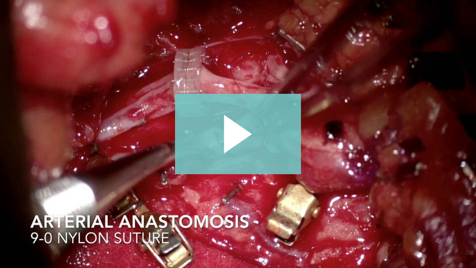
Watch the Full VideoThis video demonstrates an external dacryocystorhinostomy surgery with insertion of a nasolacrimal duct stent in a patient with a history of dacryocystitis of rare fungal etiology.
Watch the Full VideoAbstract Introduction: Eagle syndrome can affect many patients of any age, anywhere from 25 to 80 years old. The most common symptoms are ear and anterior superior neck pain underneath the angle of the jaw, tinnitus, some throat symptoms, and dizziness. There are two approaches that can be done for surgery, with our preference being for the intraoral approach. The pathophysiology is that the stylohyoid ligament becomes calcified and can cause pressure on blood vessels and nerves, causing variable symptoms. It is frequently undiagnosed causing patients to visit several physicians before correctly identifying the problem. A CT scan of the neck with or without contrast, can help identify the problem. Case presentation: A 39-year-old female with a history of ear and upper neck pain at the angle of the jaw. CT imaging showed calcification of the stylohyoid ligament. Surgery was recommended and a trans-oral approach was used. Methods: General anesthesia with muscle relaxation was used. A crow Davis or Dingman tractor was used to retract the endotracheal tube to allow exposure of the Oropharynx. Betadine was used to help sterilize the oropharynx. Palpation on each side is done to localize the calcified ligament and if present, the surgery is much easier to do. A 2.5 cm vertical incision is made in the anterior tonsillar pillar, being careful not to go too high on the soft palate because it can paralyze the soft palate causing significant reflux into the nasopharynx and nose, with speech and swallowing problems. The tonsil capsule and the medial pterygoid muscle are identified, and the dissection is between the two. The calcified ligament is usually about 2.5 cm deep to that area. It is in or under the fat pad in the prevertebral area. It may be difficult to find, and it is helpful if your finger is passed through the incision to palpate deeper to feel the bony process. The stylohyoid muscle and fat must be cleaned off the bone as high and low as can be dissected ideally using a combination of the monopolar and bipolar cautery. It is important to be careful in this area with the monopolar cautery because of the proximity to the internal carotid artery and jugular vein. Also, the vagus nerve can be injured. A Kerrison rongeur is used to fracture the bone superiorly. The ligament is connected at the inferior part which can be divided with the cautery. It is important to obtain good hemostasis using the bipolar cautery and saline irrigation. The wound is closed by sewing the tonsil capsule to the medial pterygoid muscle after which the mucosal incision is sewed. Preferably, vicryl sutures are used so that it will last approximately four weeks. Bupivacaine 0.5% can be injected around the surgical site to decrease postop pain. The surgery is done as an outpatient basis and the patient is given pain medication and antibiotics for significant throat pain lasting 7 to 10 days postop. Conclusion: The removal of the calcified Stylohyoid ligament via an intraoral approach, can be simple or very complicated, and must be done carefully by an experienced surgeon to avoid major complications. Most patients benefit significantly with relief of their symptoms and are very grateful. This case illustrates the surgical procedure that was easy to perform, but they are not all that easy. Surgeons: Siddharth Patel, MD James Y Suen, MD Conflicts of Interest: None Funding: This research received no external funding Department of Otolaryngology – Head and Neck Surgery, University of Arkansas for Medical Sciences, Little Rock, AR, USA
Watch the Full VideoVelopharyngeal insufficiency (VPI) is a speech disorder characterized by inability for the palate (velum) to contact the posterior pharyngeal wall resulting in nasal air escape and subsequent speech abnormalities. All but the mildest cases are treated surgically, with technique chosen based on the closure pattern and gap size reserving the pharyngeal flap for the most severely affected patients. We present a 6-year old female with developmental delay and VPI with large (~60%) coronal pattern velopharyngeal gap subsequently deemed a candidate for posterior pharyngeal flap. Following Dingman mouthgag placement, the posterior wall is inspected and palpated ensuring no carotid medialization. The flap is designed and marked as wide and long as possible to reduce tension. Local injection wis performed. Using an angled needle tip Bovie, the flap was then elevated in the plane the prevertebral fascia to the level of the nasopharynx. The donor site is closed with simple interrupted 4.0 chromic sutures. The palatal mucosa is divided in a T-shaped fashion, without violating palatal musculature. The flap is inset with horizontal mattress sutures using 4.0 chromic. The nasal ports are inspected frequently to ensure adequate nasal airway patency. The palatal mucosa is reapproximated and any residual donor site closed. The patient is observed overnight, discharged home post-operative day 1, maintained on a soft diet for two weeks and abstains from speech therapy for 4 weeks to allow healing. Follow up demonstrated excellent healing well and VPI resolution on repeat speech sample.
Watch the Full VideoThis video demonstrates an overview of radial arterial cannulation in a pediatric patient using real-time ultrasound (US) guidance. Ultrasound imaging is a useful tool in the armamentarium for guiding arterial line placement, and its use has become commonplace due to increased accessibility and improved technology. Ultrasound imaging facilitates the detection of abnormal anatomy and abnormal findings (e.g. thrombosis). It also allows for real-time guidance for arterial cannulation, which is especially valuable during difficult insertions such as in neonates or small infants, patients with weak or absent pulses or landmarks, and those with multiple prior cannulations. There is evidence of higher success rates with first attempts and decreased complications compared to the traditional landmark and palpation techniques.
Watch the Full Video32-month-old male with Coffin Siris syndrome, bilateral middle ear effusions, and velopharyngeal insufficiency who presents with a submucous cleft palate.
Watch the Full VideoThis video demonstrates the placement of a central venous catheter (CVC) in the internal jugular vein (IJV) in an infant using real-time ultrasound (US) guidance. Traditionally, the landmark approach has been the technique used to guide CVC placement. Presently, the use of ultrasound (US) for guiding placement has become commonplace due to increased accessibility, improved technology, and evidence of increased first-attempt success rates and decreased complications. Real-time US-guided central venous cannulation is now the recommended technique over the landmark technique by professional organizations. The experienced use of US allows for the detection of abnormal anatomy or findings (e.g. vein thrombosis) and allows for real-time visualization, which is especially helpful during difficult insertions, absence of landmarks, and in challenging patient groups such as in small infants.
Watch the Full VideoSpecialty
Plastic Surgery
Orthopedics
The Scalpel Report
Dive into the latest surgical innovations and insights
with our curated biweekly newsletter
Our Partners
CSurgeries highlights top surgical tools from our industry partners. Explore
videos featuring cutting-edge surgical tools from leading surgical industry brands.
Articles and Blogs
Modern vs. Traditional Surgical
Education: A Comparative
Analysis
August 23, 2023
The 6 Uses of Video Clips in
Modern Surgical Education
August 17, 2023
Keeping fit may help prevent atrial fibrillation and stroke
August 10, 2023
Upcoming Webinars
Advance Techniques of
Esophageal Dilation
Date: 08/05/2023 | 4 ATM
Series, Otalaryngology, Upcoming
Webinars 38
By: Peter C. Belafsky, MD, MPH, PhD
The Science Behind Vaccines
Date: 08/05/2023 | 4 ATM
Series, Otalaryngology, Upcoming
Webinars 38
By: Peter C. Belafsky, MD, MPH, PhD
An Interactive Salivary Case Study
Date: 08/05/2023 | 4 ATM
Series, Upcoming, Webinars 38
By: Peter C. Belafsky, MD, MPH, PhD