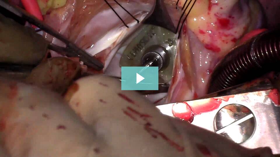
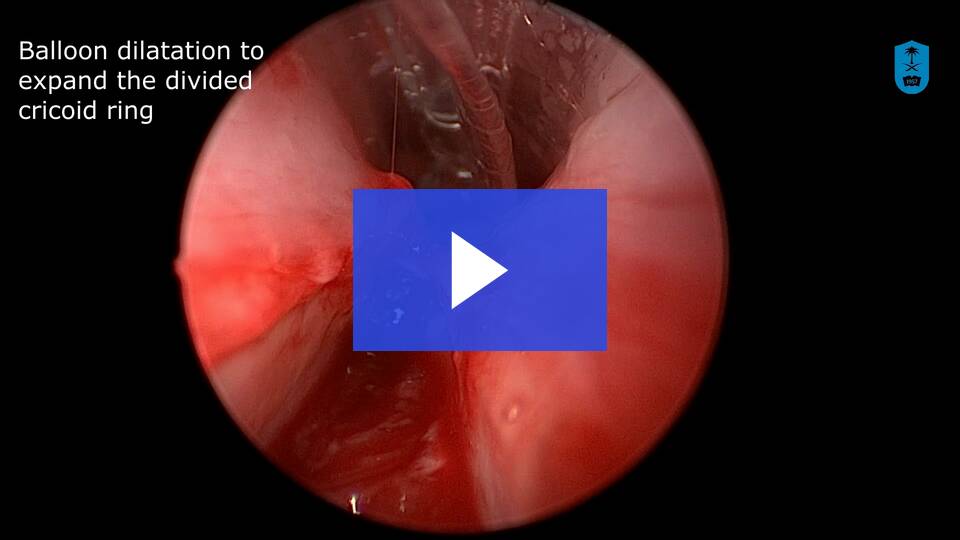
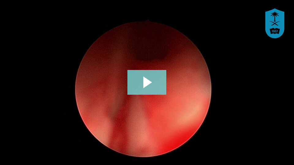
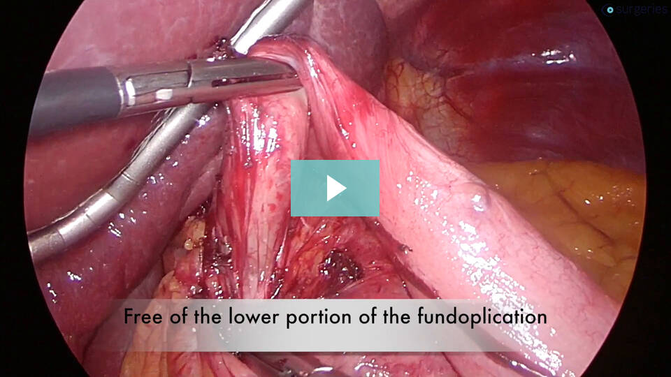
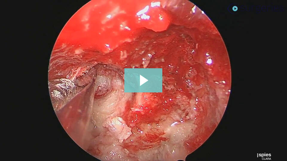
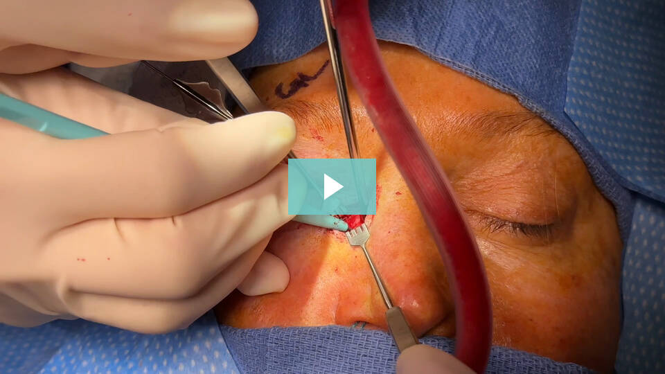
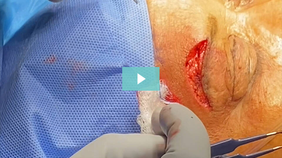
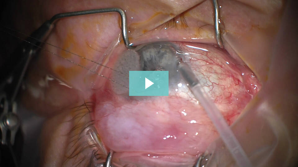
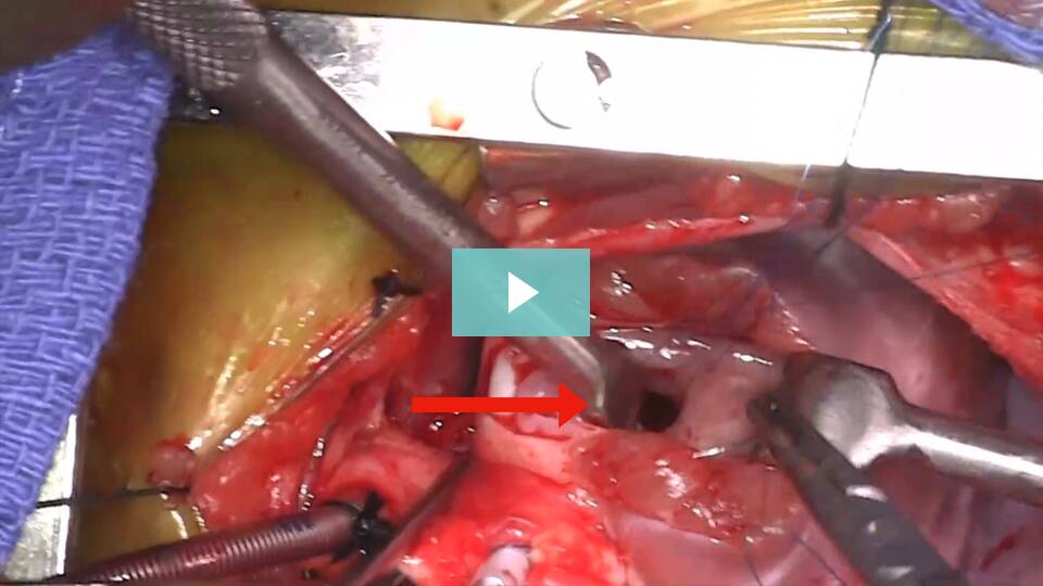
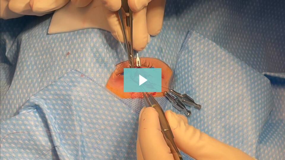
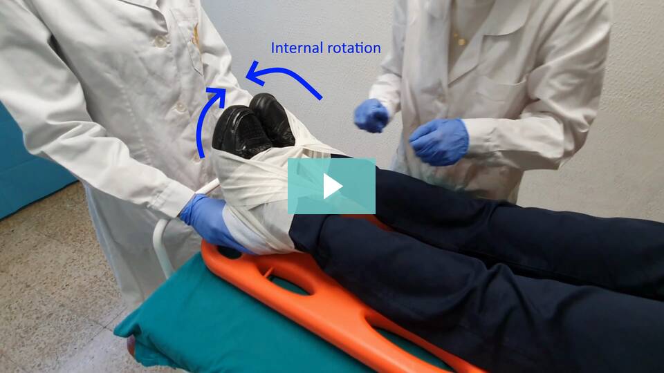
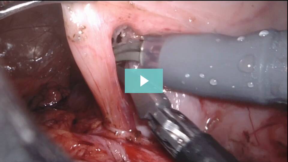
The leading global platform for
peer-reviewed surgical videos
Explore a collection of 500+ peer-reviewed surgical videos
from 300 contributors from around the world.
Recently Added
Our Latest and Most Popular Videos
This video demonstrates an epidural catheter placement on a 2-year-old, 12kg male patient presenting for left hip osteotomy. His past medical history was remarkable for congenital heart defects, bilateral congenital hip dislocations, and a sacral dimple which is sometimes associated with neurologic spinal canal abnormalities. In this case, no neurologic anatomical abnormalities were demonstrated on the neonatal spine ultrasound. The patient was placed in a left lateral decubitus position. Using anatomical landmarks like Tuffier’s line or the intercristal line corresponding to L4-L5 level, the target level for needle placement was identified and marked. The patient’s skin was sterilized and draped under sterile conditions. An 18-gauge, 5 cm length Tuohy needle was used to encounter the epidural space. A general guideline for the depth to the epidural space from the skin is approximately 1mm/kg of body weight¹. Subsequently, a 20-gauge catheter was placed through the needle to a depth of 4.5 cm at the level of the skin. Negative aspiration of blood or CSF was confirmed. A test dose was calculated at 0.5 mcg/kg epinephrine or 0.1ml/kg of lidocaine 1.5% with epinephrine 1:200,000. In this case, a 1.2 mL test dose of lidocaine 1.5% with epinephrine 1:200,000 was given without any observed cardiovascular changes (e.g. ≥ 25% increase or decrease in T wave amplitude, HR increase ≥ 10 bpm, or SBP increase ≥ 15 mmHg)¹. Finally, the catheter was secured to the back of the patient. Parental consent was obtained for the publication of this video.
Watch the Full VideoZ-plasty allows broken-line closure, reorientation of the defect in the horizontal plane with re-creation of a cervicomental angle, and most importantly, a lengthening of the anterior neck skin that aids in preventing recurrent contracture. We present our experience managing a congenital cervical midline cleft in a 3-month-old patient and describe a simple technique for planning the ideal Z-plasty closure. No simple description for planning the ideal closure for this defect could be found in the otolaryngology literature.
Watch the Full VideoObjective tinnitus is a rare phenomenon whereby a patient perceives sound in the absence of external auditory stimuli, that is also observed by the examiner. Unlike subjective tinnitus which is thought to be somatosensory and usually difficult to cure, objective tinnitus is more likely to have an identifiable cause amenable to treatment. The differential for objective tinnitus includes aberrant vascular anatomy affecting the temporal bone, patulous eustachian tube function, and abnormal myoclonic activity of the palatal or middle ear muscles.1 We present a 16-year-old female who presented for evaluation of objective tinnitus. On physical examination, an intermittent rhythmic clicking was identified. Visualization of both the tympanic membrane and palate during active audible tinnitus was observed and found to be normal. A hearing test was performed demonstrating normal hearing and speech thresholds as well as normal tympanogram. Acoustic reflex testing demonstrated absent decay in both ears and spontaneous discharge for the right ear in response to both high and very low stimulus indicating abnormal stapedial and tensor tympani function. MRA demonstrated normal vascular anatomy and MRI was obtained demonstrating normal anatomy without lesions of the brainstem, cochleovestibular nerves, or ear or mastoid pathology. The patients was subsequently diagnosed with isolated middle ear myocolonus (MEM). Treatment options including medical versus surgical therapy were discussed as has previously been described. The patient ultimately elected for surgical tenotomy of the stapedial and tensor tympani tendons. Using endoscopic technique, a middle ear exploration was performed. Canal injection was performed with standard tympanomeatal flap elevation was assisted with epinephrine pledgets. The Annular ligament was identified and the middle ear was entered. Additional dissections was performed superiorly, and the chorda tympani nerve was identified and preserved. The stapedial tendon was visualized emanating from the pyramidal eminence to the posterior crus of the stapes. Balluci scissors were used to sharply incise the tendon and the remaining ends were reflected using a Rosen needle to prevent re-anastamosis. Additional dissection along the malleus was performed to gain access to the tensor tympani tendon. A 30 degree angled endoscope was utilized to visualize the tensor tympani tendon extending forward from the cochleariform process to the neck of the malleus. The angled 6400 Beaver blade was used to sharply incise the tendon, requiring multiple passess due to the thickness of the tendon. The sharply incised ends of both tendons were clearly visualized. The tympanomeatal flap was re draped and secured with gel foam packing. The patient was seen in follow up three weeks post operatively with a well healed ear drum, resolution of her objective tinnitus, normal hearing, and absent stapedial reflexes. The patient and mother were happy. Endoscopic stapedial and tensor tympani tenotomy is a feasible technique for isolated MEM in the pediatric population.
Watch the Full VideoVenous malformations (VM) are congenital lesions, frequently affecting the head and neck, with poor respect for tissue planes. Established treatments include observation, sclerotherapy, laser, and surgical resection.1 Lesions affecting the upper airways present unique challenge due to frequent unresectability and difficult access/exposure for alternative standard treatments. We describe our approach of standard endoscopic airway techniques for the administration of advanced treatment modalities including simultaneous laser and sclerotherapy for an extensive airway VM. Our patient is a 16-year-old female with an extensive multi-spatial VM with associated airway obstruction. The patient suffered from severe obstructive sleep apnea (OSA) and continuous positive airway pressure (CPAP) dependence as a result of airway compression. Direct laryngoscopy and bronchoscopy demonstrated extensive venous staining and large vascular channels of the hypopharynx. Lumenis Nd:Yag laser (Yokneam, Israel) via 550 micron fiber was passed under telescopic visualization. Treatment via previously described “polka dot” technique was performed (15W, 0.5 pulse duration) with immediate tissue response. The largest vascular channel was accessed via 25-gauge butterfly needle. Immediate return of blood following lesion puncture confirmed intralesional placement. Reconstituted bleomycin (1 U/kg; max dose = 15 U per treatment) was injected and hemostasis achieved with afrin pledgets. The patient was intubated overnight. She was extubated the next morning and advanced to a regular diet, discharging post-operative day two. Post-operative flexible laryngoscopy demonstrated significant improvement in the treatment areas, and follow up sleep study demonstrated sleep apnea resolution with liberation of her CPAP therapy.
Watch the Full VideoA 47-year-old male, with a history of multiple cholelithiasis and multiple choledochal lithiasis, who presented with multiple episodes of cholangitis for which endoscopic treatment (ERCP + stenting) was performed. After 4 unsuccessful attempts to resolve the bile duct by endoscopic approach, it was decided to perform minimally invasive laparoscopic surgery. In this video we can observe the Choledochotomy, followed by extraction of stones and biliary mud. Subsequently, a choledochoscopy is performed with the laparoscopic camera (10 mm) with infusion of sterile Physiological Solution since the patient had a very dilated bile duct. Choledochorrhaphy is then performed.
Watch the Full VideoA 75-year-old male with history of chronic HCV- related hepatitis, in regular follow-up and sustained viral response (SVR), presented at our Emergency Department for sudden epigastric pain. Urgency CT scan and subsequent abdominal MRI revealed a 2,5cm monofocal HCC in S5 with surrounding hepatic hematoma (7cm of extension) and hemoperitoneum layer. The procedure consisted in laparoscopic exploration, lysis of tenacious adhesions between hepatic hematoma and the right colic flexure, intraoperative ultrasound to assess tumor extension, preparation of Pringle Maneuver and parenchyma transection with ultrasound dissector combined with colecistectomy.
Watch the Full VideoAcquired tracheomalacia in the form of suprastomal collapse may occur as a complication of long-term tracheotomy dependence. Prolapse of the weakened suprastomal segment of trachea during inspiration may prevent safe decannulation. Management of such an issue may require a secondary surgical procedure such as anterior tracheoplasty.2 In 2001, Forte et al described the use of thyroid ala cartilage as a reliable cartilage source for anterior augmentation laryngotracheal reconstruction in neonates. This technique may yield a favorable result given similar thickness of the cartilages and use of a single incision operation for airway reconstruction.1 Here, we present a modification of the procedure described by Forte for anterior cervical tracheoplasty for the indication of suprastomal collapse preventing decannulation. The procedure begins with nasotracheal intubation and excision of tracheostomy tract and stoma. Strap muscles are then divided to expose the laryngotracheal cartilages. Cartilages are divided at the midline anteriorly, and the diseased segment of anterior trachea is discarded. The defect is measured, and if the size match is favorable, the superior thyroid alar cartilage is harvested. The resulting cartilage graft is slightly larger than the tracheal defect and is placed so that the perichondrium is facing into the airway lumen. Interrupted sutures of 4-0 vicryl are used to inset the graft in a submucosal fashion. Once the graft is secured with sutures, a Valsalva maneuver is performed after the cuff of the endotracheal tube is taken down to assure no leak. Strap muscles are reapproximated, a Penrose drain is placed, and the skin is closed. The child is kept intubated and sedated for 3 days before subsequent extubation in the intensive care unit. A bronchoscopy is performed at the 6-week postoperative interval to assure successful healing and to remove any persistent granulation tissue if present.
Watch the Full VideoThis video demonstrates an evisceration surgery with placement of a 16mm silicone implant in a patient with a blind, painful eye.
Watch the Full VideoThis video demonstrates punctal dilation and insertion of a Mini-Monoka stent for treatment of epiphora due to punctal/canalicular stenosis.
Watch the Full VideoSpecialty
Plastic Surgery

Orthopedics


The Scalpel Report
Dive into the latest surgical innovations and insights
with our curated monthly newsletter
Our Partners
CSurgeries videos feature talents from top surgical institutions, collaborations from organizations
as well as cutting-edge tools from leading surgical industry brands from around the world.
Articles and Blogs
Latest Webinars

Advanced Techniques of Esophageal Dilation

The Ins and Outs of Medical Research & Publication

Voice Feminisation Surgery


